A Machine Learning Approach to Assess the Separation of Seismocardiographic Signals by Respiration
Executive Summary
This study investigates the use of machine learning to classify seismocardiographic (SCG) signals based on respiratory and lung volume phases. Using Support Vector Machines (SVM), Random Forests (RF), and Neural Networks (NN), the study achieved high subject-specific classification accuracies (up to 95%) but lower generalization in leave-one-subject-out scenarios. Clustering analysis using K-means demonstrated that grouping SCG signals by lung volume phases (high/low) is more effective than by respiratory phases (inspiration/expiration), with purity and adjusted rand index scores favoring lung volume separation.
Answer Machine Insights
Q: What was the primary finding regarding the grouping of SCG signals?
Grouping SCG signals by lung volume phases (high/low) was more effective than by respiratory phases (inspiration/expiration).
The clustering results when using frequency features suggests that separating SCG signals according to their lung volume phase may be superior to separating according to respiration phase.
Q: Which machine learning algorithm performed best in subject-specific classification?
All three algorithms (SVM, RF, NN) performed similarly well, achieving up to 95% accuracy.
The classification accuracy with the SS training scenarios for predicting respiration/lung volume phases when using a SVM, RF, and NN were 95%/94%, 95%/90%, and 95%/94% respectively.
Key Results
Subject-specific classification accuracy for lung volume phases reached 95%.
K-means clustering purity for lung volume phases was 0.81, compared to 0.65 for respiratory phases.
Visual Evidence
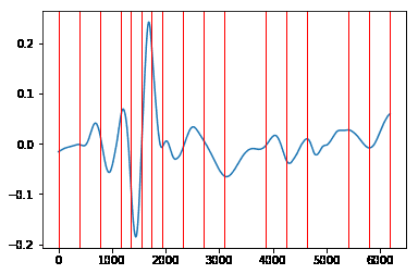
Figure 3.2: SCGz divided into 16 adaptive-width bins
Clinical Snapshot
Evidence Rating
Relevance
high Priority