Comparative analysis of three different modalities for characterization of the seismocardiogram
Executive Summary
This study compares three modalities—echocardiography, Cine-MRI, and a 3D finite element electromechanical model—for characterizing seismocardiogram (SCG) signals and correlating them with cardiac events. Experimental SCG signals were measured using a high-sensitivity accelerometer, while Cine-MRI and electromechanical modeling provided alternative methods to reconstruct SCG signals. Preliminary findings suggest that these modalities can complement echocardiography in understanding SCG morphology and its relationship to cardiac mechanics, with potential applications in simulating pathological cases such as ischemia.
Answer Machine Insights
Q: What cardiac events were identified using echocardiography?
Aortic and mitral valve opening and closure, as well as rapid systolic ejection and rapid diastolic filling.
A simultaneous M-mode echocardiogram of the subjects was acquired from the parasternal long (or short) axis for detection of aortic and mitral valve opening and closure. Continuous wave Doppler was used in an apical five chamber view for detection of the peak of rapid systolic ejection.
Q: How does Cine-MRI contribute to SCG signal reconstruction?
Cine-MRI provides 3D cardiac geometry and contraction data, enabling estimation of SCG displacement and acceleration.
Using endo-epi contours, we stacked image slices for each time frame and obtained 30 volumes representing heart geometries and contraction for a cardiac cycle. By computing derivatives of this displacement we got velocity and acceleration.
Key Results
Experimental SCG signals showed clear correlation with cardiac events such as valve opening and closure using echocardiography.
Cine-MRI and electromechanical modeling successfully reconstructed SCG signals, though Cine-MRI struggled with rapid systolic ejection and diastolic filling waves.
Visual Evidence
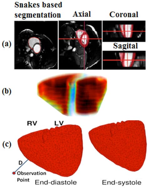
Figure 3. Cine-MRI based 3D model: a) Different views of Snakes based segmentation; b) Rough Estimation of left/right ventricle volumes in end-diastole; c) End-diastolic and end-systolic states of the ventricles obtained from cine MRI. Line labeled with D shows how we could use these deformable meshes to capture acceleration of cardiac vibrations by computing second derivative of D’s displacement changes over mesh deforms.
Clinical Snapshot
Evidence Rating
Relevance
high Priority