Investigating Seismocardiogram Patterns: A Computational Modeling of Cardiac Wall Motion Propagation to the Chest Surface
Executive Summary
This study employs finite element modeling (FEM) to simulate seismocardiogram (SCG) signals by tracking cardiac wall motion using the Lucas-Kanade optical flow algorithm applied to 4D CT images. The computational domain includes anatomical structures such as lungs, ribcage, and chest muscles, and the simulated SCG signals are validated against actual SCG data from the literature. Key cardiac events, including valve openings and closures, are identified, demonstrating the potential of FEM for understanding SCG signal genesis and improving non-invasive cardiac monitoring.
Answer Machine Insights
Q: What cardiac events were identified using the simulated SCG signals?
Mitral valve closure (MC), aortic valve opening (AO), and aortic valve closure (AC) were identified.
Important cardiac events such as mitral valve closure (MC), AC, and AO were determined based on the LV waveform.
Q: How was cardiac wall motion tracked for the FEM simulations?
Cardiac wall motion was tracked using the Lucas-Kanade optical flow algorithm applied to 4D CT scan images.
A custom MATLAB implementation of the Lucas-Kanade optical flow algorithm was developed to track the time-resolved displacements of the heart wall throughout a complete cardiac cycle.
Key Results
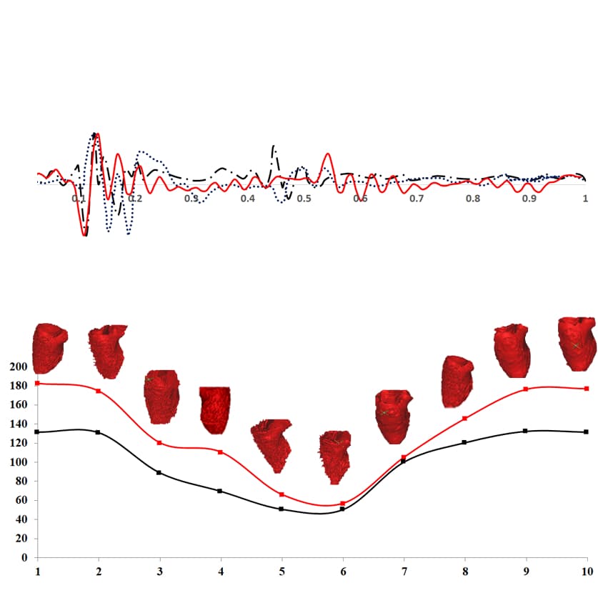
Simulated SCG signals successfully replicated fiducial points such as aortic valve opening (AO) and closure (AC).
Left ventricular volume derived from CT scans correlated well with SCG waveforms from prior studies.
Visual Evidence
Clinical Snapshot
Evidence Rating
Relevance
high Priority