Seismocardiography and 4D flow MRI reveal impact of aortic valve replacement on chest acceleration and aortic hemodynamics
Executive Summary
This study investigates the impact of aortic valve replacement (AVR) on chest vibrations and aortic hemodynamics using seismocardiography (SCG) and 4D flow MRI. Pre- and post-surgical measurements reveal that AVR resolves pathological flow patterns associated with bicuspid aortic valve disease, as evidenced by reduced high-frequency SCG energy and cohesive flow in MRI. The findings suggest SCG as a cost-effective adjunct for post-operative evaluation of valve-mediated flow pathology.
Answer Machine Insights
Q: What changes in SCG signals were observed after AVR?
Post-surgical SCG signals showed reduced high-frequency energy during systole and increased energy at valve closure.
In the second half of systole, the average high-frequency SCG energy is reduced by 46% to 0.28 mm/s2, and SCG acceleration energy at aortic close averaged over 60–240 Hz is 0.95 mm/s2, or 66% higher.
Q: How did 4D flow MRI measurements change after AVR?
MRI showed resolution of jetting and helical flow, with cohesive flow patterns and reduced peak velocity in the ascending aorta.
Pathline visualizations from 4D flow MR depict largely cohesive flow without jet patterns. Maximal peak velocity in the ascending aorta observed during systole is 161 cm/s.
Key Results
Post-surgical SCG high-frequency energy reduced by 46% during systole, indicating resolution of pathological flow.
Peak velocity in the ascending aorta decreased from 343 cm/s pre-surgery to 161 cm/s post-surgery, as measured by 4D flow MRI.
Visual Evidence
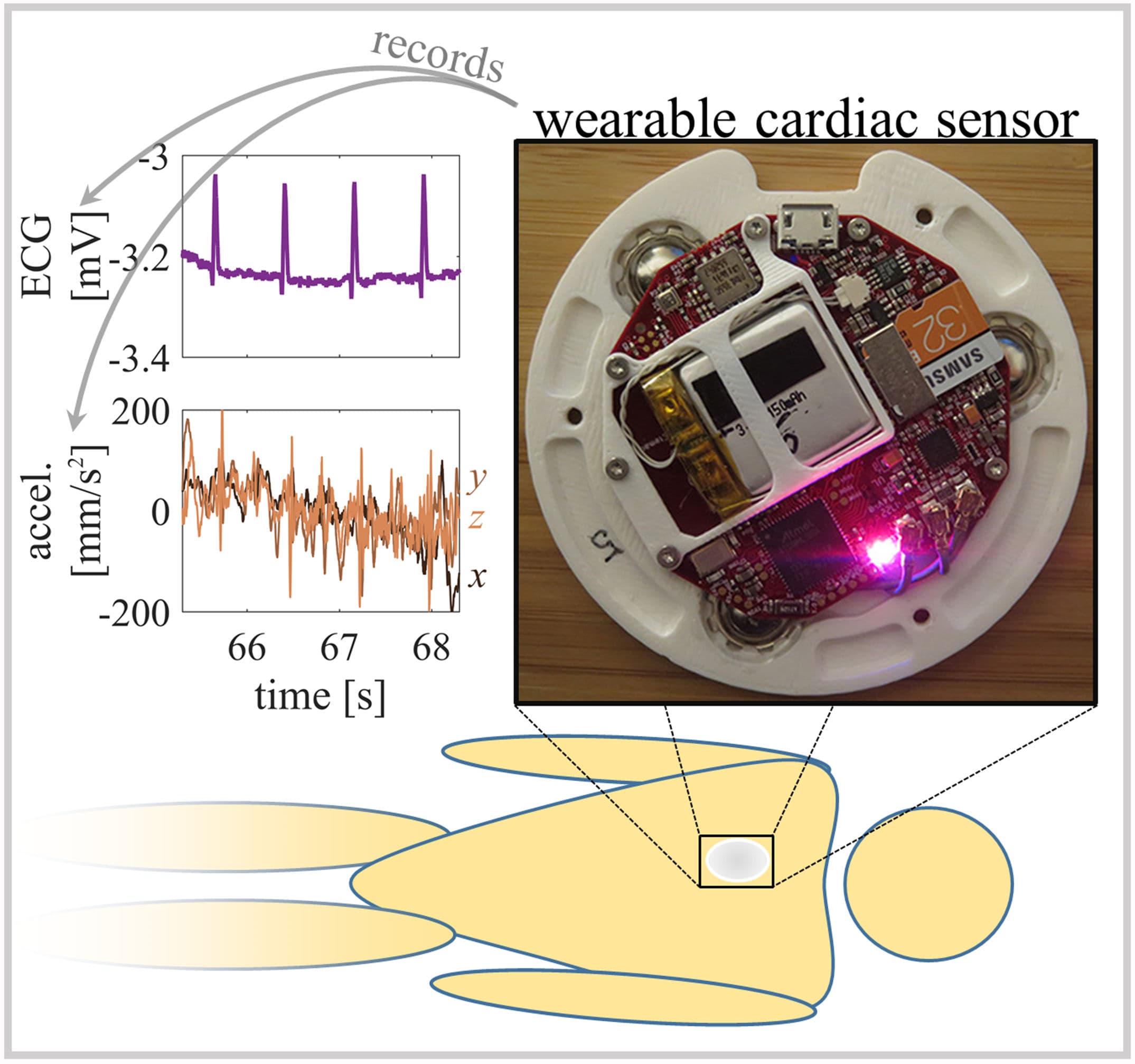
Figure 1. A custom wearable cardiac monitoring device recording ECG and chest accelerations recorded the supine SCG at the sternum.
Clinical Snapshot
Evidence Rating
Relevance
high Priority