A trimodal system for the acquisition of synchronous echocardiography, electrocardiography, and seismocardiography data
Executive Summary
This study presents a novel trimodal system for synchronous acquisition of echocardiography, electrocardiography (EKG), and seismocardiography (SCG) data. The system integrates custom hardware and software components to enable high-precision data collection and synchronization, facilitating the study of mechanical and electrical heart characteristics. A feasibility test demonstrated the system's capability to acquire synchronized data, with potential applications in cardiac imaging triggering and ventricular function monitoring.
Answer Machine Insights
Q: What is the primary goal of the trimodal system?
To evaluate the feasibility of triggering cardiac imaging techniques like CT and MRI using mechanical signals from echocardiography or SCG instead of electrical signals from EKG.
Our ultimate goal is to develop a real-time system that evaluates the mechanical motion of the heart to determine the quasi-stationary periods within a cardiac cycle during which acquisition of cross-sectional data by CT or MRI can be triggered.
Q: What hardware components were used in the custom device?
The custom device includes amplifiers, a power supply unit, a multi-channel analog-to-digital converter (ADC), and a hardware interface.
The custom-built device consists chiefly of amplifiers, a power supply unit, a multi-channel analog-to-digital converter (ADC), and a hardware interface.
Key Results
The system successfully acquired synchronized echocardiography, EKG, and SCG data from a human subject.
Preliminary analysis showed variability in SCG features relative to EKG signals between subjects, suggesting individual differences in cardiac mechanical states.
Visual Evidence
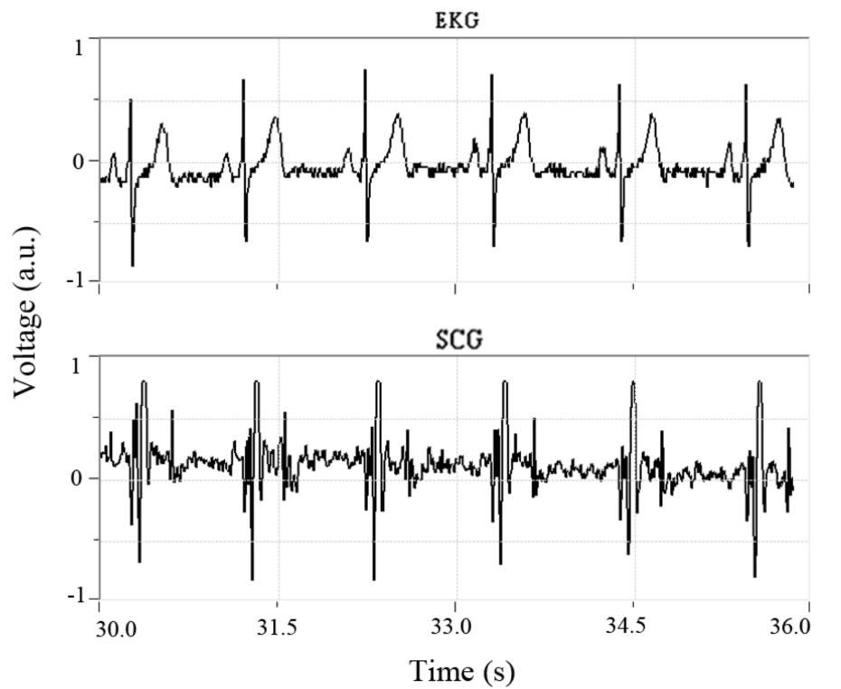
Fig. 5. Software interface for our custom device displaying the simultaneously acquired EKG data (top) and SCG data (bottom).
Clinical Snapshot
Evidence Rating
Relevance
high Priority