Mechanical deconditioning of the heart due to long-term bed rest as observed on seismocardiogram morphology
Executive Summary
This study investigates the mechanical deconditioning of the heart during prolonged 60-day head-down tilt bed rest (HDT) using seismocardiography (SCG) and functional data analysis (FDA). Key findings include significant decreases in SCG fiducial amplitudes (AO and AC), pulse transit time (PTT), and left-ventricular ejection time (LVET), indicating reduced mechanical heart strength and increased arterial stiffness. Seasonal variations were observed between campaigns, suggesting environmental influences on cardiovascular responses. The study highlights SCG's potential for monitoring cardiovascular changes in microgravity and terrestrial applications.
Answer Machine Insights
Q: What cardiovascular changes were observed during prolonged bed rest?
Significant decreases in SCG fiducial amplitudes (AO and AC), PTT, and LVET were observed, indicating reduced heart mechanical strength and increased arterial stiffness.
SCG fiducial morphology AO (aortic valve opening) and AC (aortic valve closing) amplitudes showed significant decrease between BDC12 and HDT52 (p < 0.03). PTT and LVET were also found to decrease through HDT bed rest (p < 0.01).
Q: How did seasonal differences affect cardiovascular responses?
Seasonal differences influenced blood pressure and heart rate, with campaign 1 (January) showing more drastic decreases in PTT and blood pressure compared to campaign 2 (September).
PTT decrease more drastically between pre- to post-bed rest in Campaign 1 compared to Campaign 2 (p < 0.01).
Key Results
PTT decreased by 15–40% during HDT, indicating increased arterial stiffness.
SCG fiducial amplitudes (AO and AC) showed significant decreases (p < 0.03), suggesting reduced mechanical heart strength.
Visual Evidence
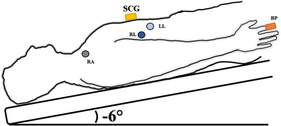
Fig. 1 HDT schematic of sensor placement. SCG (yellow rectangle) placed on the xiphoid process. Blood pressure measured at the finger (orange rectangle). ECG Lead II shown RA lead (gray circle) on right clavicle, RL lead (dark blue circle) on lower right rib cage and LL (light blue circle) on lower left rib cage.
Clinical Snapshot
Evidence Rating
Relevance
high Priority