Detecting Aortic Stenosis Using Seismocardiography and Gryocardiography Combined with Convolutional Neural Networks
Executive Summary
This study investigates the use of seismocardiography (SCG) and gyrocardiography (GCG) signals combined with convolutional neural networks (CNNs) to detect aortic stenosis (AS). Using a custom data logger and continuous wavelet transform for signal processing, the model achieved high specificity (98.42%), sensitivity (98.14%), and accuracy (98.36%) in distinguishing AS patients from healthy controls. The findings suggest a promising non-invasive diagnostic tool for AS, potentially reducing reliance on echocardiography performed by specialists.
Answer Machine Insights
Q: What is the primary diagnostic performance of the proposed method?
The method achieved specificity of 98.42%, sensitivity of 98.14%, and accuracy of 98.36%.
Using leave-subject-out cross validation, the model produced specificity of 98.42%, sensitivity of 98.14%, and average accuracy of 98.36%.
Q: What signal processing techniques were used to prepare the data?
Continuous wavelet transform (CWT) was applied to produce time-frequency representations of cardiac cycles, followed by dynamic time warping (DTW) for beat selection.
The saved cardiac cycles were then processed using a continuous wavelet transform (CWT). The output of the CWT produced a time-frequency matrix representation of each cardiac cycle.
Key Results
Specificity of 98.42%, sensitivity of 98.14%, and accuracy of 98.36% in detecting aortic stenosis.
The CNN model trained on SCG and GCG signals outperformed similar studies in classification metrics.
Visual Evidence
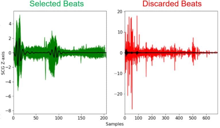
Figure 2: Overlaid selected and discarded cardiac cycles from a subject diagnosed with AS. The plotted Z-axis SCG
Clinical Snapshot
Evidence Rating
Relevance
high Priority