Robustness of Persistence Diagrams to Time-Delay for Seismocardiogram Signal Quality Assessment*
Executive Summary
This study evaluates the robustness of topological data analysis (TDA) using persistence diagrams for SCG signal quality assessment under time-delay conditions. It compares TDA with dynamic time feature matching (DTFM) and demonstrates that TDA significantly outperforms DTFM in ranking SCG beats by signal-to-noise ratio (SNR) when beats are segmented earlier than ECG R-peak locations. These findings highlight the potential of TDA for ECG-free SCG signal quality analysis, enabling longitudinal cardiac monitoring in ambulatory and critical care settings.
Answer Machine Insights
Q: How does TDA perform compared to DTFM under time-delay conditions?
TDA significantly outperforms DTFM in ranking SCG beats by SNR under time-delay conditions.
For all shifted cases (100, 200, and 300 ms), p<0.001 and post-hoc analyses showed significant differences between all pairs of models.
Q: What is the practical advantage of TDA for SCG signal quality assessment?
TDA does not require fine-tuning of hyperparameters, making it more user-friendly and robust for ECG-free SCG signal quality analysis.
TDA does not require fine-tuning thus decreasing the burden on the user.
Key Results
TDA maintained a mean Kendall's Tau value above 0.94 across all time-delay conditions, demonstrating robust ranking performance.
DTFM performance dropped significantly under time-delay conditions, with Kendall's Tau values becoming negative for DTFMD and diminishing to 0-0.4 for DTFMS.
Visual Evidence
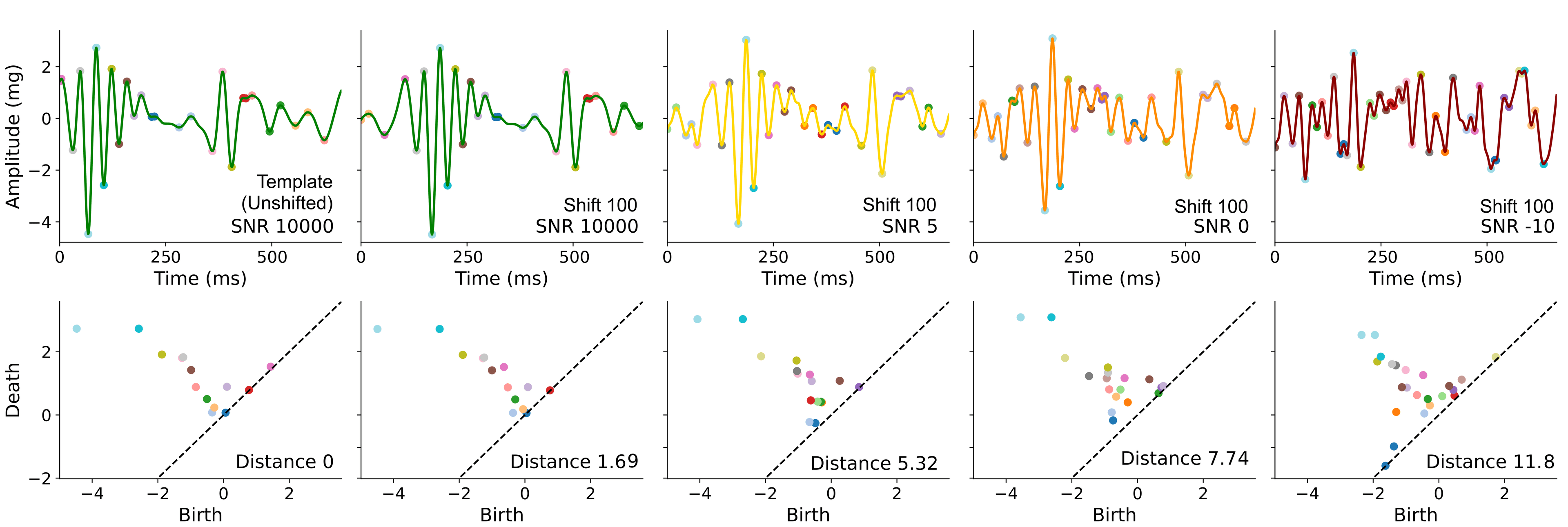
Fig. 2. Example seismocardiogram beat traces and corresponding persistence diagrams (PDs): We extracted PDs for the unshifted template beat and beats containing varying levels of added synthetic noise at multiple shift amounts. Beats of four SNR levels (10000, 5, 0, and -10) which were segmented 100 ms earlier than the ECG R-peak are shown above with decreasing SNR. Peaks and valleys are colored in accordance with their matching points on the PDs.
Clinical Snapshot
Evidence Rating
Relevance
high Priority