Executive Summary
This study introduces a novel adaptive recursive least squares filter (ARLSF) for motion artifact removal in seismocardiography (SCG) signals, utilizing a single consumer-grade accelerometer. Tested on 16 subjects during standing and walking conditions, the ARLSF achieved a heartbeat detection accuracy of 98% and demonstrated strong agreement between SCG-derived and ECG-derived heart rates. The method shows promise for improving SCG signal clarity without additional processing, paving the way for wearable applications in dynamic environments.
Answer Machine Insights
Q: What is the accuracy of heartbeat detection using ARLSF?
The heartbeat detection accuracy using ARLSF was up to 98%.
The heartbeat detection accuracy was up to 98% and heart rates estimated from the SCG and ECG matched well under both the standing and walking conditions.
Q: How does the heart rate estimated from SCG compare to ECG?
The heart rates estimated from SCG and ECG matched very well, with a mean difference of 0.08 bpm and a standard deviation of 2.08 bpm.
The detailed difference between the heart rates from ECG and SCG with a mean value of 0.08 bpm and a standard deviation value of 2.08 bpm is plotted in Figure 12.
Key Results
Heartbeat detection accuracy of 98% achieved using ARLSF.
Heart rate differences between SCG and ECG had a mean of 0.08 bpm and a standard deviation of 2.08 bpm.
Visual Evidence
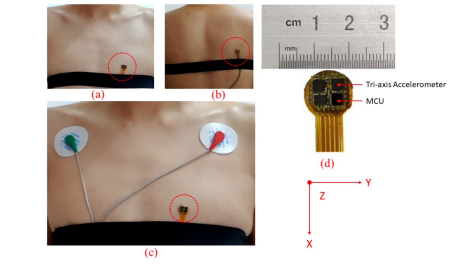
Figure 3. (a,b) Seismocardiography (SCG) recorder system placement for measuring heartbeat signal and motion signals, respectively. (c) A pair of electrocardiogram (ECG) lead I electrodes placement and SCG recorder system placement. (d) An image of the SCG recorder system which shows the dimensions. X-axis, y-axis and z-axis describe the head to foot, shoulder to shoulder and dorsoventral direction, respectively.
Research Tags
Clinical Snapshot
Evidence Rating
Relevance
high Priority