A new algorithm for segmentation of cardiac quiescent phases and cardiac time intervals using seismocardiography
Executive Summary
This study developed an algorithm for automatic detection of cardiac events and segmentation of cardiac quiescent phases using seismocardiography (SCG) synchronized with electrocardiography (ECG). The algorithm accurately measured systolic time intervals (STIs) such as QS2, PEP, and LVET, and identified quiescent phases critical for applications like nuclear imaging and diastolic timed vibrators. Results showed poor correlation between QS2 and heart rate but highlighted strong inverse relationships between heart rate and diastolic quiescent phases, suggesting SCG's potential for non-invasive cardiac mechanics monitoring.
Answer Machine Insights
Q: What correlation was observed between heart rate and diastolic quiescent phase duration?
A high inverse correlation (r = -0.97) was observed.
High inverse correlation (r= -0.97) was obtained between the heart rate and the diastolic quiescent phase (see Fig.5 (a)).
Q: What are the clinical applications of cardiac quiescent phase segmentation?
Applications include nuclear medicine imaging (e.g., CT or PET) and diastolic timed vibrators (DTV).
Two quiescent phases were detected which are clinically significant in order to use for diagnostic or treatment tools such as nuclear medicine imaging (e.g. for computed tomography (CT) or positron emission tomography (PET)) and diastolic timed vibrators (DTV).
Key Results
Mean corrected QS2i, PEPi, and LVETi values were 513.4±9.3ms, 111.8±6.5ms, and 401.6±8.3ms respectively.
High inverse correlation (r = -0.97) was observed between heart rate and diastolic quiescent phase duration.
Visual Evidence
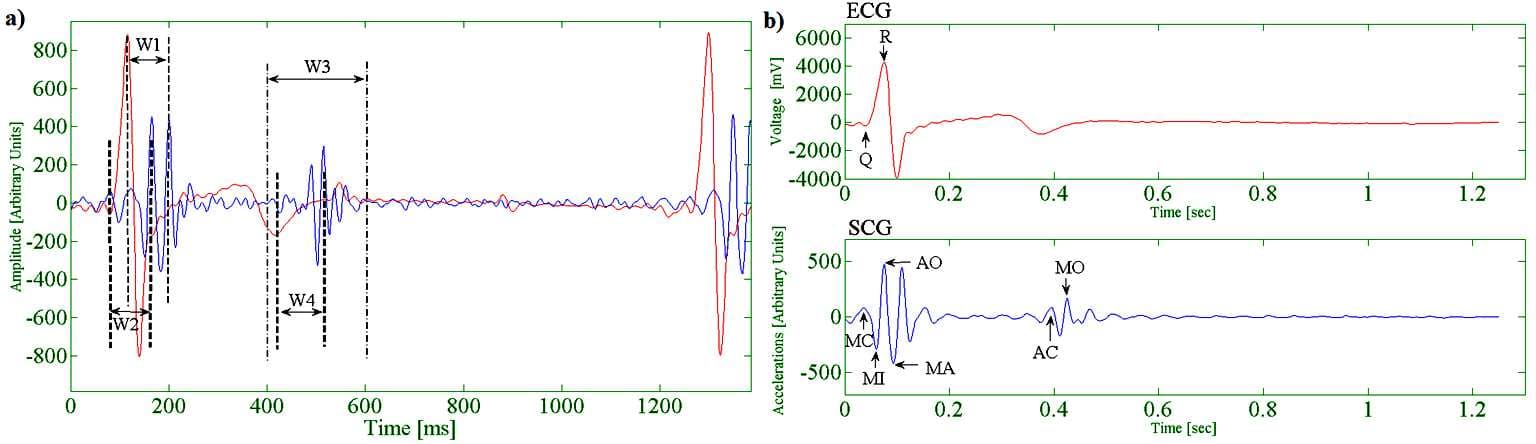
Figure 2. (a) Windowing for Event Detection. (b) Detected Cardiac Events within SCG and ECG.
Clinical Snapshot
Evidence Rating
Relevance
high Priority